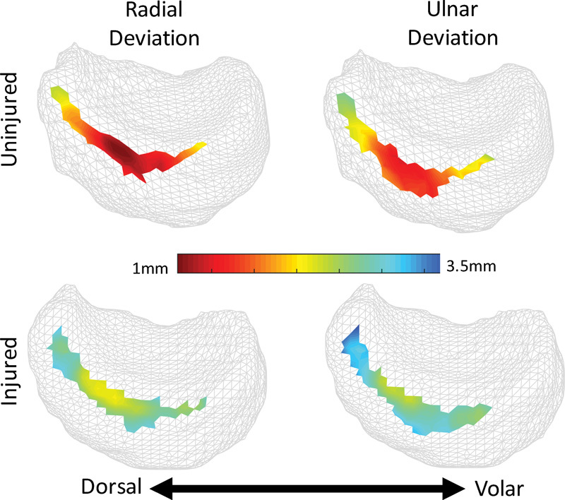
Fig. 3.

(Case 2) Sagittal view of the articular surface of the lunate. Color corresponds to interosseous distances at radial-ulnar deviation extrema during dynamic CT imaging. The injured side (bottom row) exhibits uniform, though moderate diastasis. Note: patient right-side lunate mirrored for comparison purposes.
