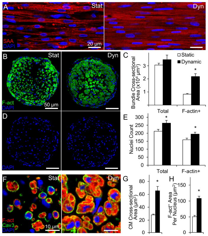
Figure 1. Effects of static and dynamic culture on structure of engineered NRVM cardiobundles.
(A) Representative images of cardiobundles cultured for 2 weeks under static (Stat) or dynamic (Dyn) conditions, stained for sarcomeric α-actinin (SAA, red) and nuclei (DAPI, blue). (B) Representative cardiobundle cross-sections stained for filamentous actin (F-act, green) and nuclei (blue). (C) Total and F-actin+ area of cardiobundle cross-sections (n = 8–10 cardiobundles per group from 4 cell isolations). (D) Cross-section stains from (B) without F-act shown. (E) Count of total and F-actin+ nuclei per cross-section (n = 8–10 cardiobundles per group from 4 cell isolations). (F) Higher magnification images of cross-sections stained for F-actin (red), Caveolin-3 (Cav3, green), and nuclei (blue). (G) Cross-sectional area of CMs (n = 4 cardiobundles per group from 2 cell isolations, with >25 CMs analyzed per cardiobundle). (H) F-actin+ area per nucleus (n = 8–10 cardiobundles per group from 4 cell isolations) *, p < 0.05 vs. static.

