
Figure 7.
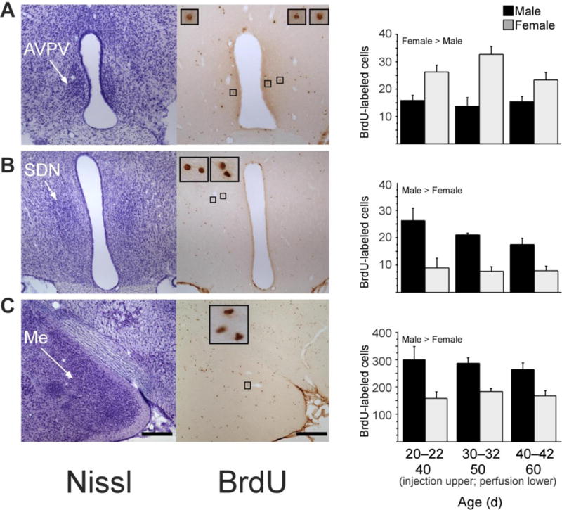
New cells are added during puberty to the AVPV, SDN and medial amygdala in male and female rats. Left photomicrographs, thionin-stained sections; right photomicrographs, BrdU labeled cells in nearby sections from the same rat; insets, BrdU-labeled cells framed in small boxes at x10 higher magnification. Rats received a daily injection of 300 mg per kg body weight of BrdU on three consecutive days at either 20–22, 30–32 or 40–42 d of age (n ¼ 6–8 per age and sex). BrdU is incorporated into DNA during the S phase of the cell cycle and can be later visualized to identify cells replicating at the time of BrdU administration. Brain tissue was collected 20 d after the first BrdU injection, on 40, 50 or 60 d of age, respectively. Quantitative analyses of BrdU-labeled cells revealed that during puberty, significantly more cells were added to AVPV (A) in females than in males, whereas significantly more cells were added to SDN (B) and medial amygdala (Me; C) in males than in females. Data are means ± s.e.m. Scale bars, 250 mm in lower-magnification images. Adapted from Ahmed et al., (2008), Nature Neuroscience 11 (9) 995–997.