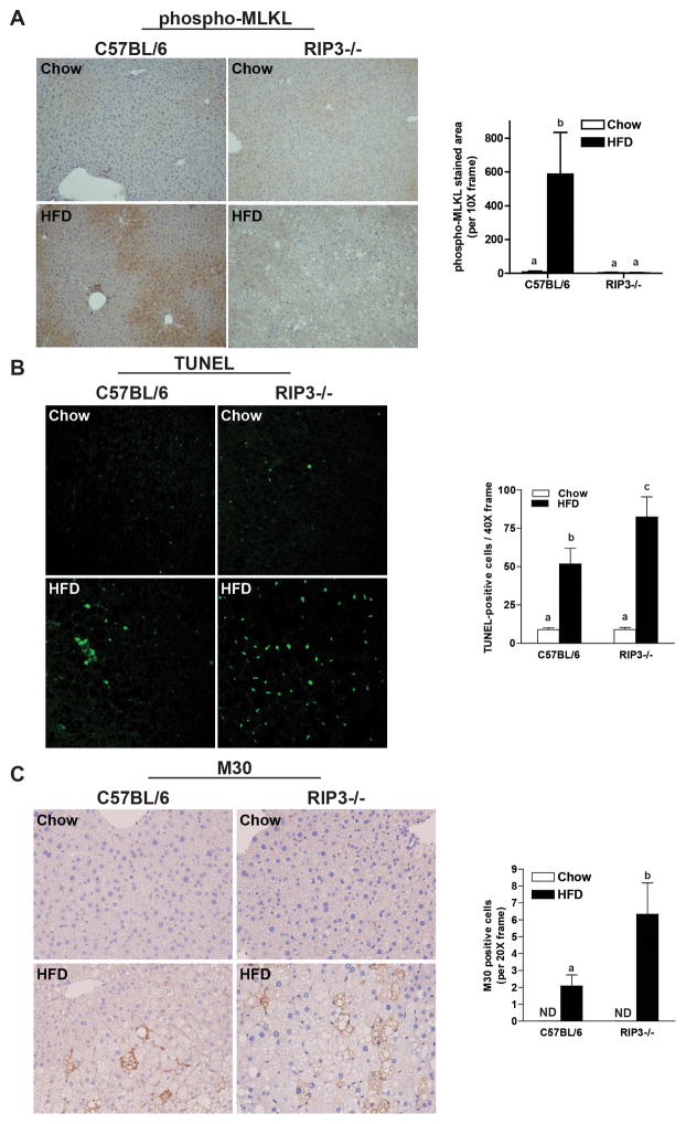
Figure 6. RIP3-deficiency decreases phosphorylation of MLKL, but promotes apoptosis in the liver in response to high-fat diet feeding.
Male C57BL/6 and RIP3-deficient mice (RIP3−/− neo) were fed HFD or chow for 12 weeks. Paraffin-embedded livers were de-paraffinized followed by (A) phospho-MLKL, (B) TUNEL or (C) M30 staining. Images were acquired using (A) a 10X objective for phospho-MLKL; (B) a 40X objective and TUNEL-positive cells were counted and expressed as percent positive of total number of cells per 40X frame and (C) a 20X objective and M30-positive cells were counted and expressed as total number of cells per 20X frame. ND: M30-positive cells were not detectable in livers of chow-fed mice. *p< 0.05 indicates a difference between genotypes in the HFD groups.

