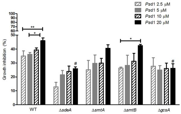
Figure 10.
Psd1 inhibitory activity depends partially on GlcCer synthesis and fungal sphingoid base structure. Wild-type, ΔsdeA, ΔsmtA, ΔsmtB and ΔgcsA cells were grown in YUU medium containing 2.5 (hatched grey bars), 5 (filled grey bars), 10 (hatched black bars) and 20 μM Psd1 (filled black bars) until OD540≥ 1.0. The growth inhibition in defensin-treated cultures was calculated using as parameters the suspensions maintained in the absence of antifungal drugs (0 % inhibition) or in 10 μM itraconazole (100 % inhibition). The values represent the means ± SEM, n = 3; *p < 0.05, 20 μM-treated WT versus 5 μM-treated WT and 20 μM-treated ΔsmtB versus 2.5 μM-treated ΔsmtB; **p < 0.01, 20 μM-treated WT versus 2.5 μM-treated WT; and #p < 0.001, 20 μM-treated ΔsdeA versus 20 μM-treated WT and 20 μM-treated ΔgcsA versus 20 μM-treated WT.

