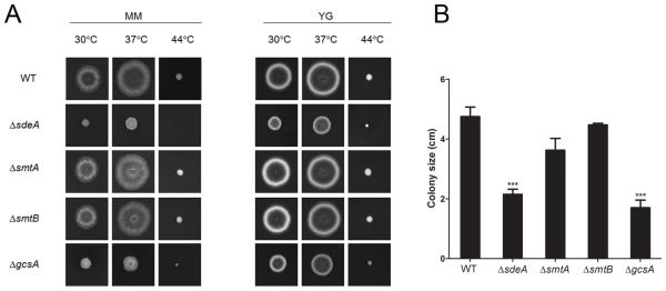
Figure 3.
GlcCer formation and sphingoid base structural modifications contribute to A. nidulans hyphal growth and differentiation. The ΔsdeA and ΔgcsA mutants showed reduced growth compared to ΔsmtA, ΔsmtB and wild-type strains. (A) Conidial suspensions of WT, ΔsdeA, ΔsmtA, ΔsmtB and ΔgcsA strains were spotted in minimal (MM) or complete (YG) solid media supplemented with uracil, uridine and pyridoxine and grown at 30, 37 or 44 °C for 2 days. (B) The colony size was determined through growth diameter measurement at 37 °C. The results are the average of three repetitions ± SEM; ***p < 0.001.

