
Figure 2.
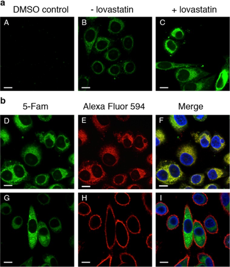
Imaging of prenylated proteins in mammalian cells via confocal microscopy. This indicates that a majority of C15Alk-labeled proteins localize in the endomembrane including the ER. Upon metabolic labeling (specified for each panel), cells were fixed and permeabilized. After several rinses, the cells were subjected to the CuAAC for 1 h with 5-Fam-PEG-N3 and imaged using a 60× objective. The green channel shows prenylated proteins “clicked” to 5-Fam-PEG-N3, and the blue channel shows the cell nucleus stained with Hoechst 34580. (A) Control reaction in which HeLa cells were treated with DMSO only. (B) HeLa cells were treated with 10 μM C15Alk for 24 h in the absence of lovastatin pretreatment. (C–I) HeLa cells treated overnight with 25 μM lovastatin, followed by 10 μM C15Alk for 24 h. (E) Red channel showing staining of ER by ER Tracker Red. (F) Overlay of images from D and E, along with nuclear stain (blue), showing significant colocalization of the green fluorescence with the ER. A zoomed in view of a region from this image is provided in Figure S4. (H) Red channel showing staining of PM by Wheat Germ Agglutinin conjugated to Alexa Fluor 594. (I) Overlay of images from G and H, along with nuclear stain (blue), indicate that the majority of the green fluorescence does not colocalize with PM. Size bar represents 10 μm.