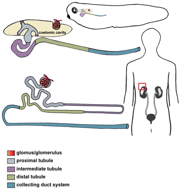
Fig. 1.
Xenopus pronephric and human metanephric nephrons share a conserved segmentation pattern. A schematic representation of an enlarged Xenopus pronephric nephron (top) and mammalian metanephric nephron (bottom), with the distinct tubular compartments labeled. The glomus/glomerulus filters the fluid from the blood plasma through capillary walls into the proximal tubule. The drawing depicts a slight difference in organization of the glomar/glomerular anatomy between the amphibian pronephric and mammalian metanephric nephrons. In mammalian metanephric nephrons, the glomerulus is integrated into the Bowman’s capsule near the proximal tubule, while the Xenopus glomus projects into a body cavity (coelomic cavity). Three branches of pronephric tubule (nephrostomes) use ciliary action to force the glomar filtrate down the pronephric tubule. Together, the tubule segments (proximal, intermediate and distal) and the collecting duct system (connecting tubule and duct) facilitate the process of filtrate reabsorption and transport of wastes products for secretion

