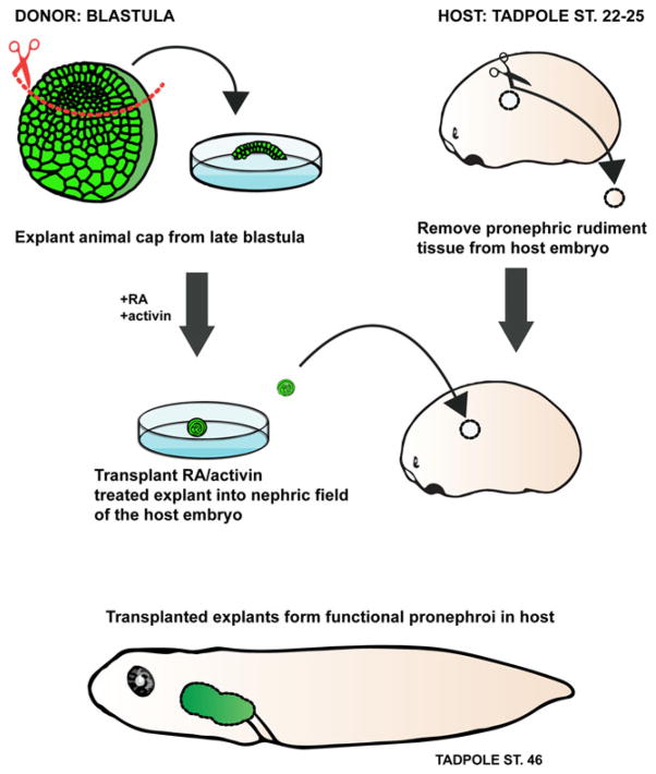
Fig. 2.
Xenopus in vitro-induced kidney organoids. Isolated animal cap (presumptive ectodermal tissue) explants (green) treated with activin and retinoic acid (RA) differentiate to form pronephric organoids in culture. The animal cap tissue treated with activin and RA can be transplanted into a host embryo which lacks kidney primordia. The in vitro-induced pronephric tissue is capable of functioning in vivo and compensates for the loss of the host kidneys by suppressing edema formation

