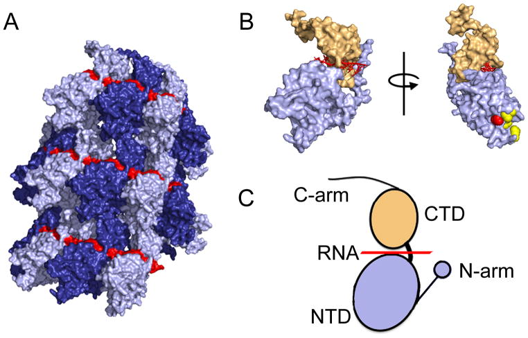
Figure 4.
Structural models of the RSV N protein and the nucleocapsid. (A) Structure of the helical RSV nucleocapsid showing the encapsidated RNA in red and individual N protein protomers in dark and light blue. (B) Surface representation of a RSV N monomer (left). Rotation of the N monomer reveals the RSV P protein binding site (red sphere) and resistance mutations against RSV604 (yellow). (C) The RSV N protein is composed of two core domains, NTD (blue) and CTD (tan), which enclose the viral RNA genome (red). Terminal extensions from the NTD and CTD domains, the N- and C-arm, respectively, interact with adjacent N subunits in the helical nucleocapsid structure. Images of protein structures were generated in Pymol. PDB codes:4BKK(RSV N), 4UCB(RSV NTD-P).

