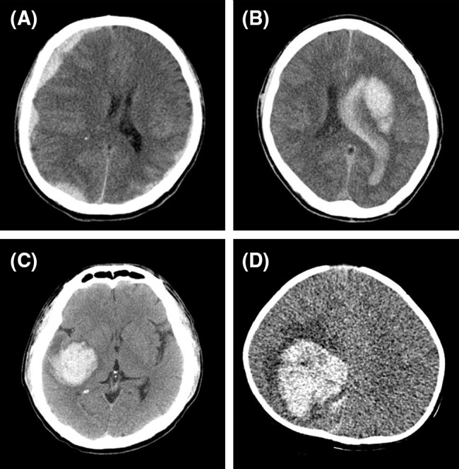
Fig. 1.

Brain computed tomography (CT) scans in four patients with hemophilia presenting with intracranial hemorrhage. a A 39-year-old man with severe hemophilia A presented with subdural hemorrhage (SDH) and a GCS score of 14. Brain CT showed SDH along both cerebral convexities and the right falx. This was accompanied by the finding that the midline was shifted to the left side. b A 42-year-old man with severe hemophilia A presented with intracranial hemorrhage and a GCS score of 3. Brain CT showed an acute intracerebral hemorrhage of 6.9 × 3.2 × 5.0 cm at the left basal ganglia, an intraventricular hemorrhage with hydrocephalus, severe brain edema, and a subfalcine herniation. c A 38-year-old man with severe hemophilia B presented with ICH and a GCS score of 3. Acute intracerebral hemorrhage was present in the right temporal lobe. d A 7-month-old boy with severe hemophilia A presented with ICH and a GCS score of 9. Brain CT showed an acute intracerebral hemorrhage of 5.0 × 5.0 × 4.0 cm at the right basal ganglia and an intraventricular hemorrhage at the right parietal lobe. Three of these patients (b, c, and d) with intracerebral hemorrhage underwent neurosurgical intervention
