
FIG 4.
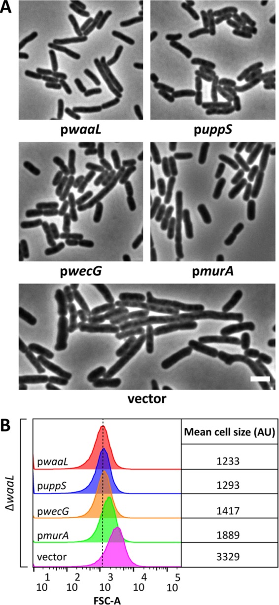
Suppression of ΔwaaL shape defects. (A) Micrographs of ΔwaaL cells containing derivatives of pDSW361 that express the indicated genes. Cells were grown at 37°C in LB containing 100 μM IPTG (0 μM IPTG for pwecG) until the culture reached an OD600 of 0.5 to 0.6. The cells were then fixed and photographed by phase-contrast microscopy. Bar, 3 μm. Further characterization of wecG overexpression is found in Fig. S3 in the supplemental material. (B) Flow cytometry data from live cells in panel A. Histograms of the forward scatter area from 100,000 events (cells) are shown. The mean cell size for ΔwaaL cells expressing waaL in trans (red graph) is represented by the dashed line and is expressed in arbitrary units (AU). Data are representative of those from two independent experiments. The strains tested were MAJ434 (pwaaL), MAJ437 (puppS), MAJ436 (pwecG), MAJ435 (pmurA), and MAJ433 (vector). The effect of expression of the aforementioned derivatives of pDSW361 on wild-type cells is shown in Fig. S4.