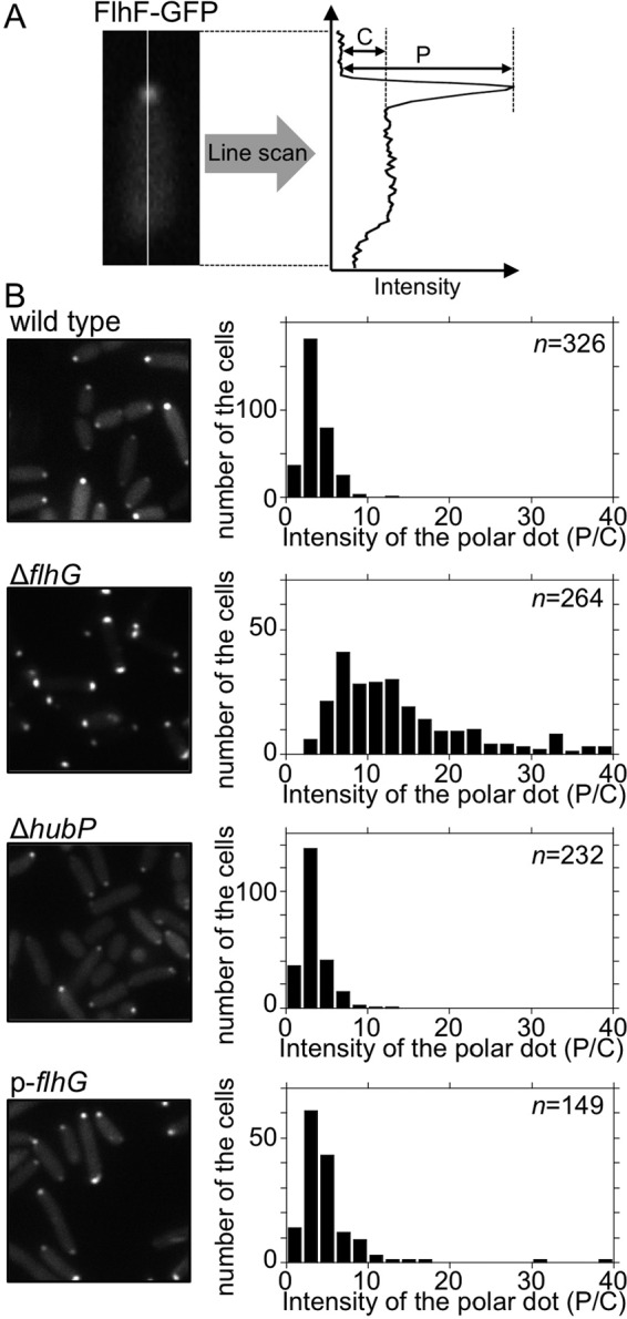
FIG 4.

Effects of hubP deletion or FlhG overproduction on the polar localization of FlhF. (A) Schematic of the quantitative analysis of the polar localization of FlhF-GFP. The fluorescence intensities at the pole (P) and in the cytoplasm (C) were determined by subtracting the background intensity. (B) Intracellular localization of FlhF-GFP and intensity of fluorescent dots. FlhF-GFP was expressed in ΔflhF cells (wild type), ΔflhF ΔflhG cells (ΔflhG), ΔflhF ΔhubP cells (ΔhubP), and ΔflhF ΔflhG cells overproducing FlhG from the plasmid pAK520 (p-flhG). Left, fluorescence images; right, P/C ratio histograms.
