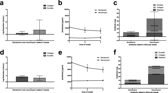
FIG 5.
Growth inhibition of MDRSA after incubation with vancomycin- and gentamicin-loaded calcium sulfate beads for 72 h and 7 days. (a) Log reductions in bacterial cell counts after 72 h of incubation of MDRSA in the model followed by a further 72 h of incubation with calcium sulfate beads loaded with vancomycin and gentamicin. Data show the decrease in viable organisms after exposure to antibiotics relative to numbers of viable organisms on exposure to unloaded control beads. (b) Corresponding concentration of antibiotics in area 1 of the models shown in panel a underneath the void, to which beads are added, through to area 3 at the edge of the model. (c) Mass of antibiotics detected in the collagen and medium phase of the models shown in panel a. (d) Log reductions in bacterial cell counts after 72 h of incubation of MDRSA in the model followed by a further 7 days of incubation with calcium sulfate beads loaded with vancomycin and gentamicin. Counts from models exposed to antibiotics are subtracted from counts for unloaded control beads. (e) Corresponding concentration of antibiotics in area 1 of the models shown in panel d underneath the void, to which beads are added, through to area 3 at the edge of the model. (f) Mass of antibiotics detected in the collagen and medium phase of the models shown in panel d.

