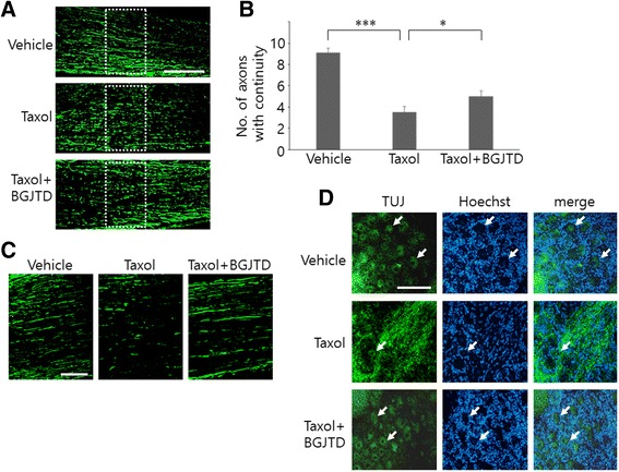
Fig. 2.

Morphological changes of sciatic nerve axons and DRG neurons after taxol injection and BGJTD treatments. Sciatic nerve was exposed and treated with DMSO vehicle, taxol, or taxol plus BGJTD, and longitudinal sections of the nerve at the injected location (a, c) and DRG at lumbar level 5 (d) were used for immunofluorescence staining for NF-200 and βIII-tubulin. The number of axons whose length is longer than 100 μm, as illustrated with dotted rectangles in (a), was counted from the images, averaged from 3 nonconsecutive sections and compared among 3 experimental groups. Quantitative data are shown in (b). Soma DRG neurons was marked by arrows in (d). *p < 0.05, ***p < 0.001 (One-way ANOVA, number of animals = 4). Scale bars in (a, c, d): 100 μm
