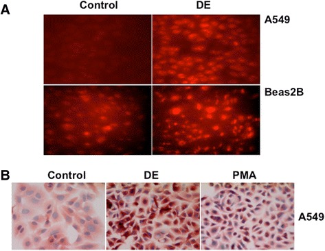
Fig. 5.

Dust extracts induce intracellular reactive oxygen species levels. a A549 and Beas2B cells grown on coverslips were incubated with 10 μM dihydroethidium for 1 h in the dark in serum-free medium and then exposed to medium alone (Control) or medium containing dust extract (1 %) (DE) for 10 min. After exposure, slides were viewed under a fluorescent microscope equipped with an Ultra-VIEW LCI scanning confocal system using 488 nm excitation and 568 nm emission filters. Fluorescent images are representative of three independent experiments. Red color indicates intracellular ROS production. b A549 cells grown on coverslips were treated with medium alone (Control), dust extract (DE) (1 %) or phorbol myristate acetate (PMA) (10 nM) for 1 h under serum-free conditions. Afterwards, hydroxynonenal conjugated proteins were visualized by immunostaining. Images shown are representative of two independent experiments. PMA was used as a positive control for the generation of intracellular reactive oxygen species (ROS). Magnification, 40×
