Fig. 1.

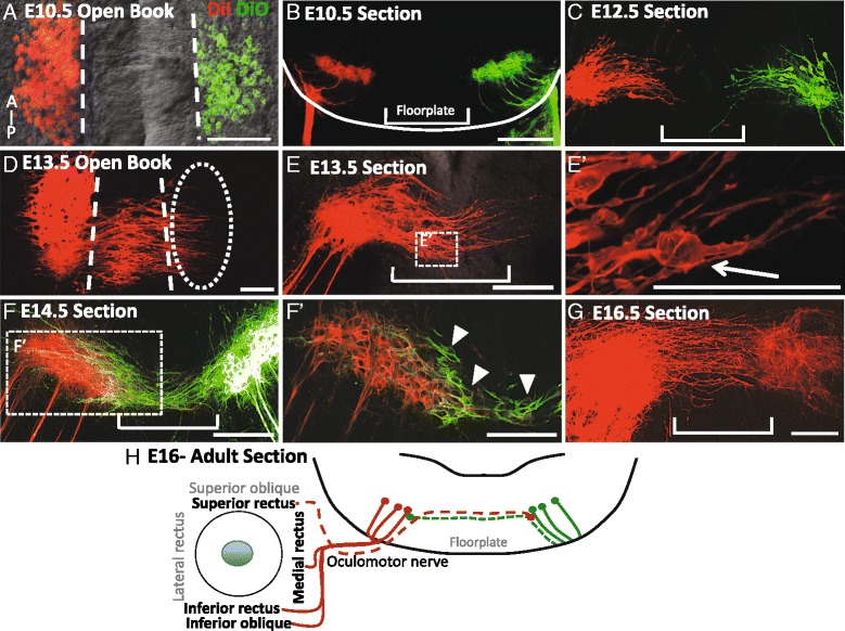
The oculomotor commissure is generated from E12.5 to E16.5 in the ventral midbrain. Oculomotor nuclei were back-labeled with peripheral application of DiI to the right oculomotor nerve (red) and DiO to the left oculomotor nerve (green) in mouse embryos on E10.5-16.5. The labeling is shown as either open book preparations revealing the anterior–posterior length of the oculomotor nucleus (A, D) or transverse sections of the midbrain (B, C, E, F, G), A, B. On E10.5, all oculomotor cell bodies were located on either side of the floor plate (A), ipsilateral to their nerve (B). C On E12.5, leading processes projected into the floor plate. D On E13.5, leading processes projected from the posterior half of the oculomotor nuclei across the midline toward the contralateral oculomotor nucleus. E Apparent cell bodies were located within the numerous leading processes within the floor plate (E’). F On E14.5, leading processes have crossed the floor plate to contact the contralateral nucleus. F’. In single focal planes by microscopy, contralateral cell bodies were located on the ventromedial aspect of the opposing nucleus as well as in the floor plate (arrow heads point to cell bodies outlined in green). G On E16.5 no cell bodies were located in the floor plate, and leading processes spanned the contralateral nucleus. H Schematic showing that the superior rectus extraocular muscle is innervated by contralateral oculomotor neurons and their midline axon fibers (dashed lines). Scale bars, 100 μm
