Fig. 3.

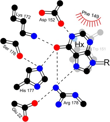
Model of ITPase substrate binding pocket. CPK coloring. Hx, hypoxanthine base of inosine; R, sugar (ribose) ring. Dashed lines indicate putative hydrogen bonds. Image reproduced from reference [31] with permission

Model of ITPase substrate binding pocket. CPK coloring. Hx, hypoxanthine base of inosine; R, sugar (ribose) ring. Dashed lines indicate putative hydrogen bonds. Image reproduced from reference [31] with permission