Figure 3.

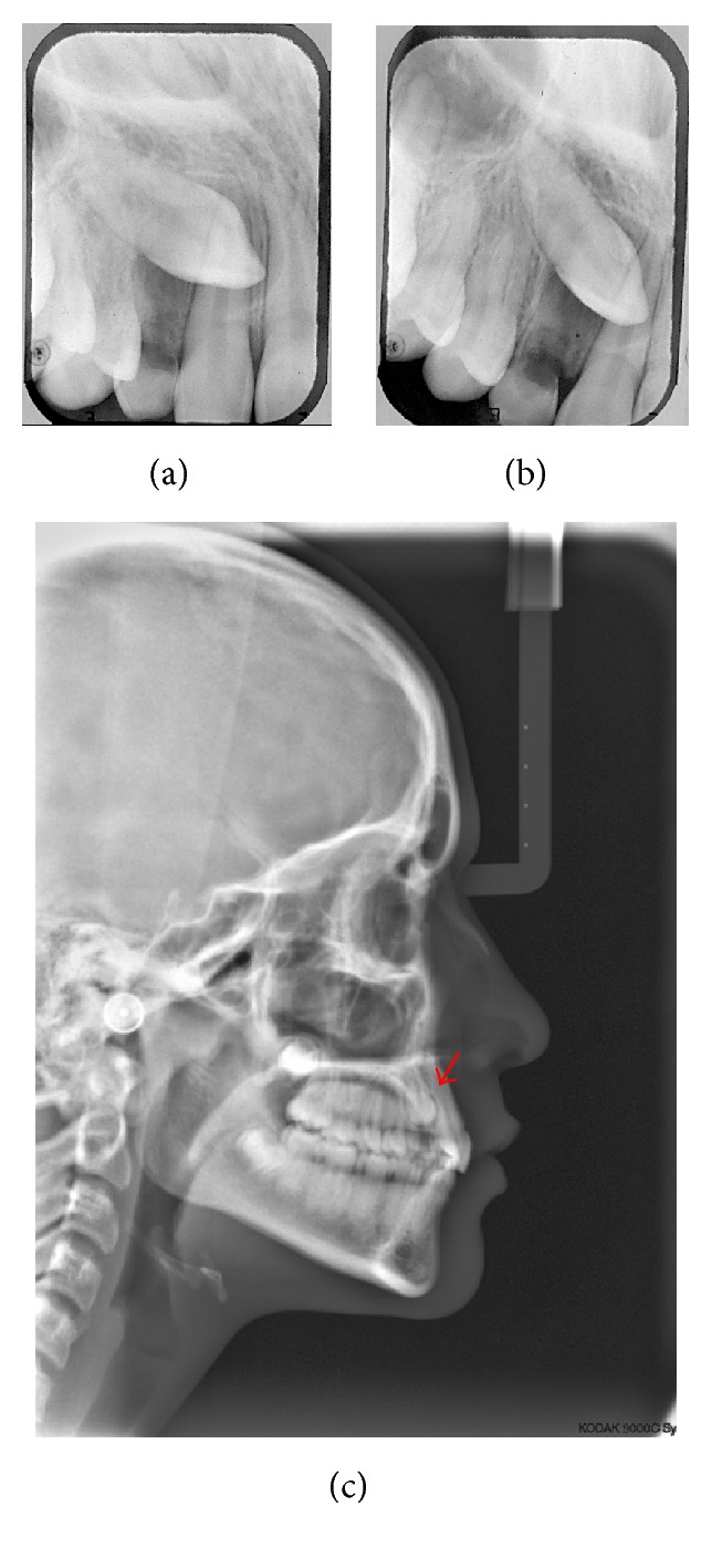
Initial documentation: exams imaging for diagnosis of impacted canine. (a) and (b) are periapical radiographs with Clark technique (note that in (b) the tooth moved distally, indicating its position by palatal); red arrow indicates the impacted tooth (c).
