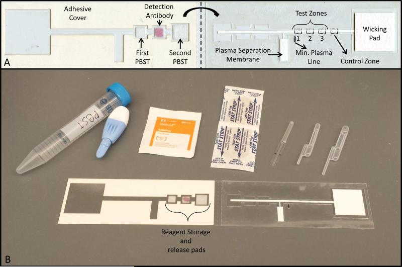
Figure 1. Two-dimensional paper network to detect human antibodies against HPV 16.
(A) Device overview. The device consists of a nitrocellulose membrane with HPV16 virus-like particles immobilized at various concentrations at three test zones (1, 2, and 3), a cellulose wicking pad on the right and a plasma separation membrane, and three glass fiber pads, one of which contains dried detection antibody, on the left. All are adhered to a thin acetate sheet. The dotted line indicates where the device is folded to start the flow of reagents through capillary action (B) All supplies needed to perform the assay at the point-of care. The supplies consist of the two-dimensional paper network device, 15 mL of phosphate-buffered saline with 0.05% Tween-20, an alcohol prep pad, a Band-Aid, a high-flow lancet, a 20 μL microsafe capillary tube and a 20μL and 40 μL exact volume transfer pipette

