
Figure 2.
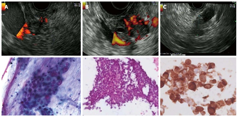
Endoscopic ultrasound and cytology images from various lesions. A: Adenocarcinoma of the pancreatic head. Cytology shows a cluster of carcinoma cells with crowding and nuclear pleomorphism with anisokaryosis and prominent nucleoli (Pap test); B: Neuroendocrine tumors (insulinoma) of the pancreas Cytology shows a group of small monomorphic cells with uniform round nuclei and fine chromatin; C: Pancreatic metastasis of malignant melanoma: Cytology shows pleomorphic tumor cells with anisokaryosis and a high nuclear-to-cytoplasmic ratio. Positive staining for melanoma antigen recognized by T-cells-1.