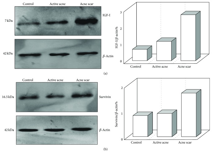
Figure 3.
Expression of IGF-I (a) and survivin (b) in the skin tissue homogenates as achieved by Western blotting. β-Actin was used in parallel as internal control. The right panels represent corresponding quantification of each gel analysis measured by Image J software and expressed as a β-actin ratio, showing higher lesional expressions of IGF-1 and survivin in the acne scar group than in the active acne group and the control group, and also lesional expressions of IGF-I in the active acne group were higher than in the control group.

