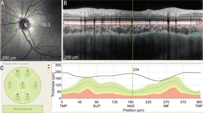
Figure 1. Example of PPCT measurements (left eye).
A: The green circle was the circular scan of a semidiameter of 1.7 mm around the optic nerve head; B: CT was measured manually from the outer part of the hyperreflective line corresponding to the base of RPE (line coloured red) to the inn*er margin of the sclera (line coloured green); C: Using the the calipers supplied by the Spectral OCT analysis software provided by Heidelberg Eye Explorer software, OCT measured the PPCT in six sectors and the globalthickness.

