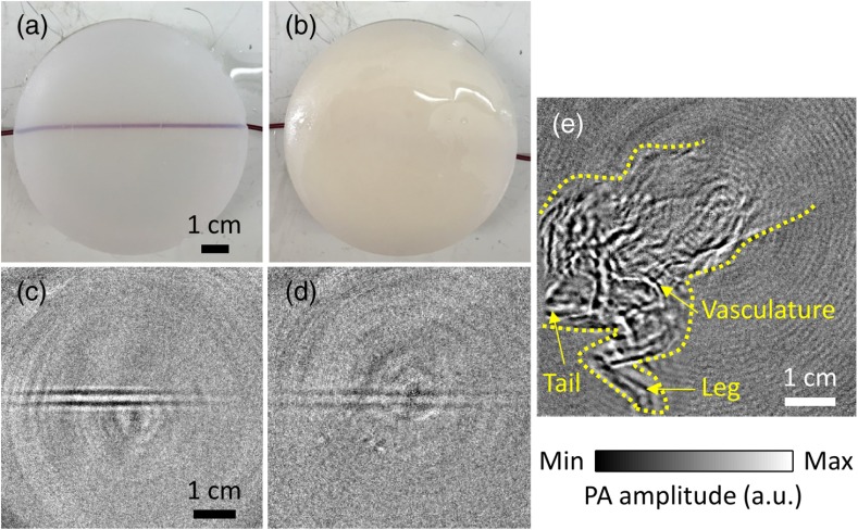
Fig. 6.
Xenon flash-lamp-based PACT of blood-filled tube phantoms made by embedding blood-filled tubes at a depth of 1 cm inside an agar–water gel without and with a scattering medium, and a whole mouse body in-vivo. (a) and (b) Photographs of the blood-filled tube phantoms without and with 1% intralipid, respectively. (c) and (d) PACT images of the phantoms (a) and (b), respectively. (e) PACT image of the whole mouse body in vivo. Different structures, such as the vasculature, leg, and tail are labeled. The mouse body is also outlined with yellow dashed lines.

