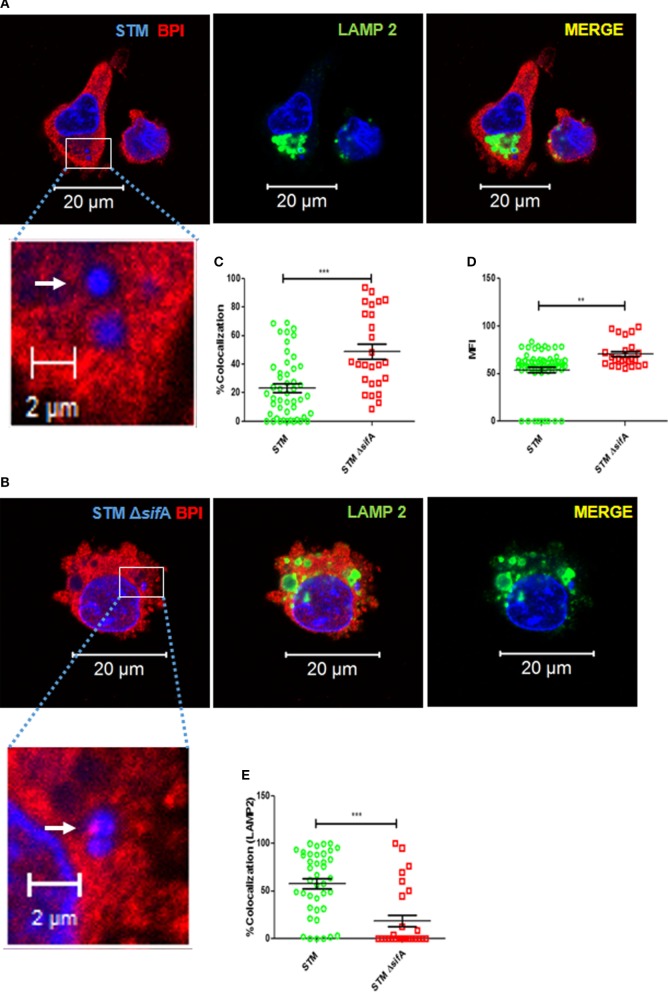
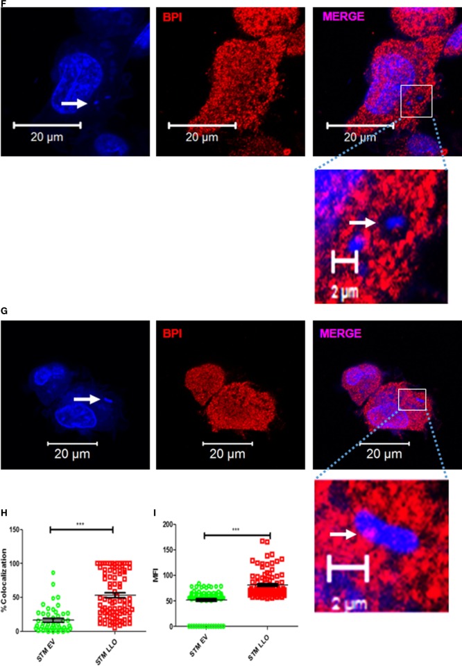
Figure 7.
STM avoids interaction with BPI inside macrophages by maintaining an actively replicating niche (SCV). U937 macrophages were infected with (A) STM, (B) STM ΔsifA, (F) STM EV (empty vector), or (G) STM LLO at an MOI of 50. Two hours post-infection, the cells were fixed with PFA at the indicated time points and stained for BPI (red) and LAMP2 (green). Nuclei and bacteria were labeled with 4′,6-diamidino-2-phenylindole (DAPI) (blue). White arrows indicate DAPI-positive bacteria. The boxed area in each set is magnified to view BPI around the bacteria. (C,H) Quantification of colocalization of BPI with bacteria. (D,I) Quantification of MFI of BPI at ROI. (E) Quantification of colocalization of LAMP2 with bacteria at ROI. All images were quantified by using the Zen Blue edition software provided by Zeiss [n = 3 (SD)]. Key: ***p < 0.001, **p < 0.005.


