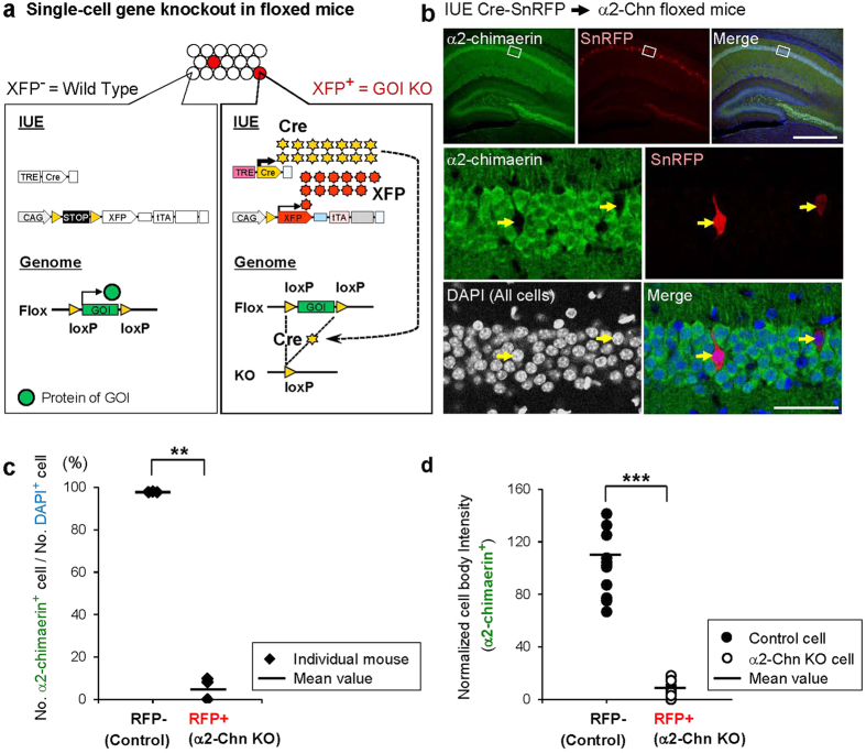
Figure 4. Labeled cell-specific gene knockout via Cre-based Supernova in floxed mice.
(a) Schematic for Supernova-mediated single-cell knockout (KO) of endogenous gene of interest (GOI) flanked by two loxP sites. (b) α2-Chimaerin protein is expressed ubiquitously in the hippocampal CA1, while it is undetected specifically in SnRFP-labeled neurons (arrows), indicating that Cre-based Supernova-mediated gene knockout is highly specific to the labeled cells. Cre-SnRFP vectors were introduced into α2-chimaerin (α2-Chn)flox/flox mouse CA1 by IUE. α2-chimaerin immunohistochemistry and DAPI-staining were performed. Upper panels show the hippocampus of α2-Chnflox/flox mouse. A set of enlarged example images is shown in the bottom. Note that because extremely high intensity signal of Supernova labeling hinders detection of α2-chimaerin signal, we partially photobleached SnRFP signal in this experiment. (c,d) Quantification of Supernova-dependent α2-Chn knockout efficiency and specificity. (c) α2-Chimaerin expression was detected in almost all of SnRFP-negative CA1 hippocampal cells (97.7% ± 0.1%, n = 3 mice; 785 cells/804 cells, 464 cells/474 cells, 1011 cells/1036 cells), while only in 5.9% ± 3.0% (n = 3 mice; 3 cells/31 cells, 0 cell/18 cells, 3 cells/37 cells) of SnRFP-positive CA1 neurons, indicating labeled cell-specific knockout. All values represent as mean ± SEM; (**): 0.001 < P < 0.01; Welch’s t-test. (d) Intensities of α2-chimaerin signal in RFP+ cells (α2-Chn KO cells) and RFP- cells (control cells) that surround RFP+ cells were measured (n = 14 cells, 3 mice for each group). (***) P < 0.001, Welch’s t-test. Scale bars: 500 μm (b, upper); 100 μm (b, bottom).

