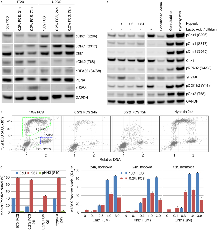
Figure 1. Modulation of the cell culture environment does not dramatically alter DDR signalling in tumour cells growing anchorage dependently.
(a) HT29 or U2OS cells were grown in low serum for 24 or 72 hours. (b) HT29 cells, in 10% FCS containing media, were grown in: hypoxia for 24 hours with or without re-oxygenation for 6 (+6) or 24 (+24) hours or normoxia with 25 mM lactic acid, conditioned media, 50 nM gemcitabine or 2.5 mM hydroxyurea for 24 hours. Cell lysates were immunoblotted using the indicated antibodies. (c) The cell cycle distribution of HT29 cells cultured as described was determined by single cell immunofluorescent analysis (n = 4, mean ± SD). (d) The fraction of HT29 cells staining positive for EdU, Ki67 or pHH3 (S10) was determined by single cell immunofluorescent analysis (n = 4, mean ± SD). (e) HT29 cells were grown under the indicated culture conditions in combination with Chk1i for 24 or 72 hours. The number of γH2AX positive nuclei was determined by single cell immunofluorescent imaging (n = 4, mean ± SD).

