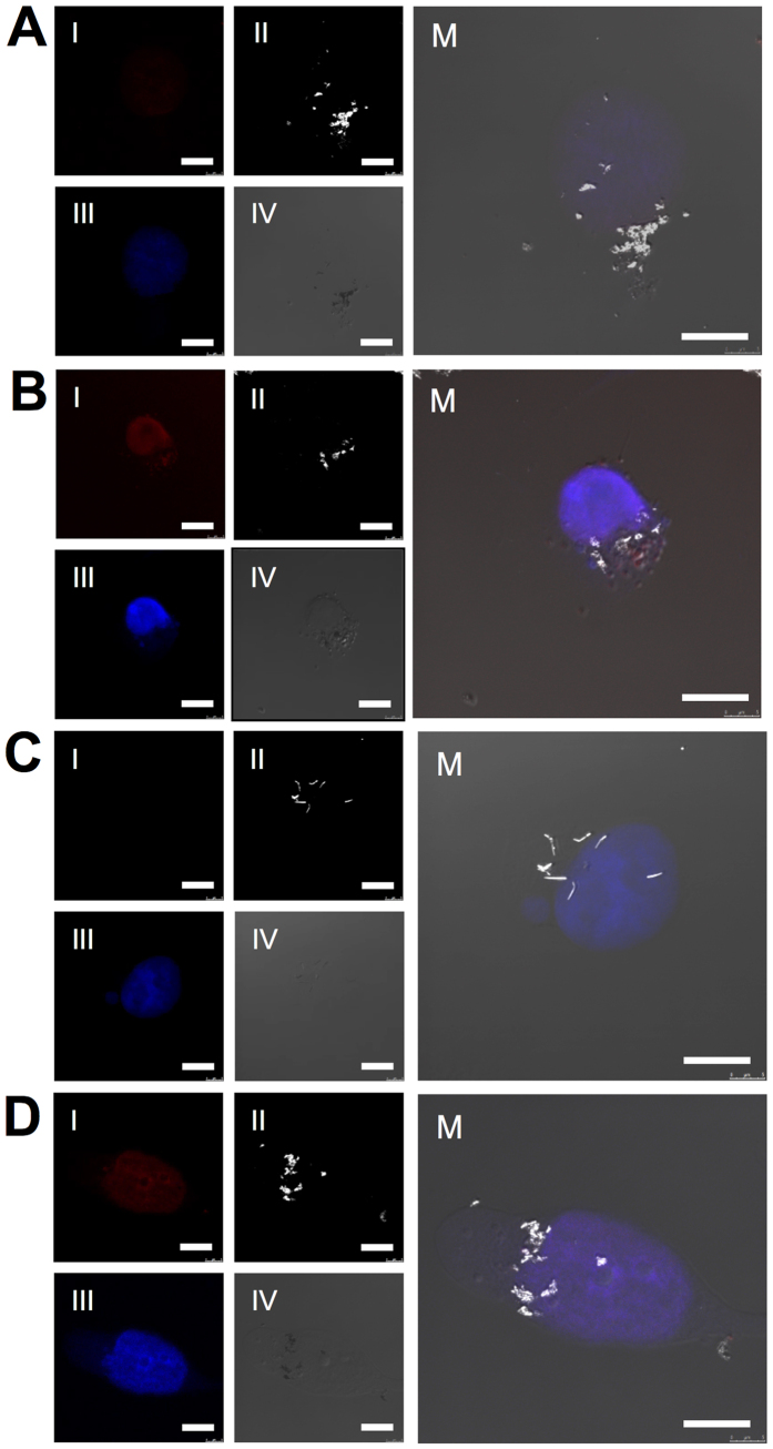
Figure 5.
Confocal microscopy images from MDA-MB-231 cells incubated with APTES-NWs-DOX after 24 h (A) and after 72 h (B) and incubated with BSA-NWs-DOX after 24 h (C) and after 72 h (D). Four different channels: I. Red fluorescence of DOX, II. Light reflected from the Fe NWs, III. DAPI nuclear staining and IV. DIC. M. shows the merged image. Scale bars = 10 μm.

