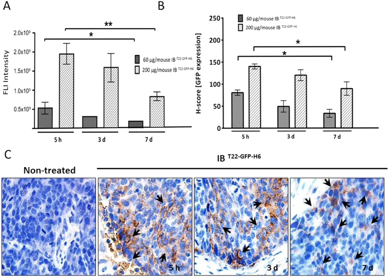
Figure 4. Fluorescent emission by IBT22-GFP-H6 deposits remaining in CXCR4+ colorectal tumors after their administration.
(A) Total IBT22-GFP-H6 protein deposits were quantified measuring fluorescent intensity at 5h, 3 and 7 days after intratumoral injection of 200 μg/mouse. Data were expressed in Radiant Efficiency. (B) Quantitation of total IBT22-GFP-H6 protein deposits plus released soluble proteins in tumor tissue calculated as an H-score for anti-GFP immunostaining (brown colour) at 5 h, and 3 or 7 days post administration. (C) Representative microphotographs of GFP inmunohistochemistry in IBT22-GFP-H6 treated tumors at 5 h, and 3 or 7 days. Note the higher intensity of GFP staining in some tumor areas at 5 h and 3 days (black arrows) and the higher dispersion of protein distribution observed inside the tumor at day 7 post-injection. Quantitative data were expressed as mean ± SE *,**Statistically significant at p < 0.05 or p < 0.01, respectively.

