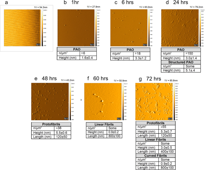
Figure 2. PAO nucleation on primary mica surface.
PAO adsorbed on primary (Iry) mica surface followed over time. Nucleation of PAO leads to the formation of linear protofibrils after 48 hrs (e). Small fibrils were also observed after 60 hrs (f) and at 72 hrs (g). Scale bars: 100 nm (f) and 200 nm (a–e,g). (a) Contact Mode (CM) topography image of starting bare mica. (b–g) Tapping Mode (TM) topography images of PAO (b–d) and protofibrils/fibrils (e–g) adsorbed on mica after different contact times (1–72 hrs) with a 44 μM Aβ42 peptide solution in 10 mM PBS at pH 7.4, T = 25 °C. The number and size of the structures are indicated in the table under each experimental time point. The amplitude scale is in Volt; we indicated the height (nm) corresponding to 1 Volt.

