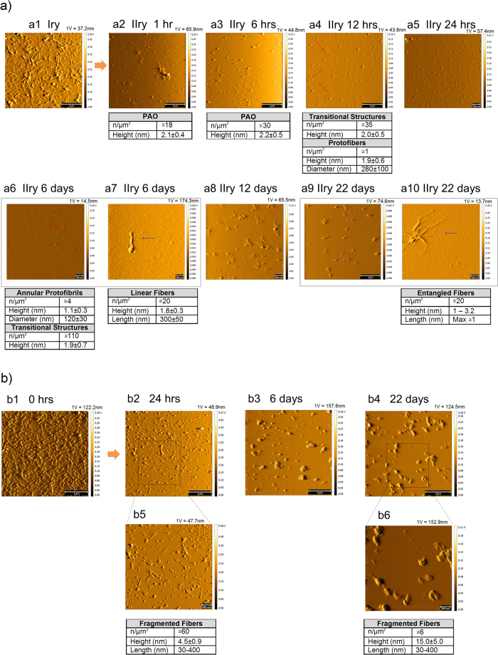
Figure 6. PAO budding and metastasis.
(a) Fibrils were adsorbed on primary (Iry) surface (scale bar: 1 μm). Empty secondary (IIry) mica was placed in semi-contact with the primary mica and followed up to 22 days. Contact solution: 10 mM PBS at pH 7.4, T = 25 °C. (a1,b1) show the TM topography image of a pristine primary coated with fibrils. Here also numerous PAO appear on the secondary surface after 1 hr whose number increases at 6 hrs. Complex structures (PAO aggregates/transitional structures and annular protofibrils – red arrows) appear as soon as 12 hrs growing in number and complexity at later time points. Scarce simple fibrils (blue arrow) appear after 6 days and more complex fibrillar structures (blue arrow) after 22 days (scale bars: 100 nm (a5–a7,a10), 200 nm (a8), and 1 μm (a2–a4, a9)). (b) TM topography images of the structure present on the primary after semi-contact at different times (1–22 days), the images b2–b6 show a progressive fragmentation and reorganization of fibers with time (scale bars: 200 nm (b5,b6) and 1 μm (b2–b4)). The number and size of the structures are indicated in the table under each experimental time point. The amplitude scale is in Volt; we indicated the height (nm) corresponding to 1 Volt.

