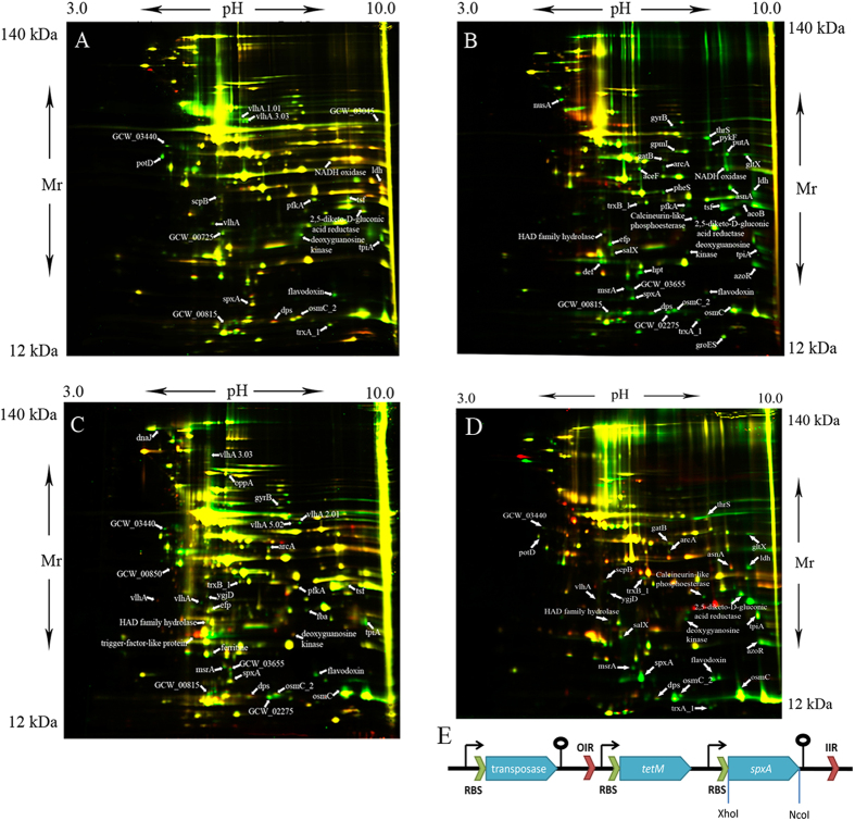
Figure 2. 2-D DIGE analysis of M. gallisepticum isolated from HD3 cells and overexpressed spxA.
(A) M. gallisepticum after 24 h of infection (Cy3, green) and laboratory strain M. gallisepticum S6 (Cy5, red). (B) M. gallisepticum after chronic infection (19 days) (Cy3, green) and laboratory strain M. gallisepticum S6 (Cy5, red). (C) M. gallisepticum after chronic infection (7weeks) (Cy3, green) and laboratory strain M. gallisepticum S6 (Cy5, red). (D) SpxA-overexpressing M. gallisepticum (Cy3, green) and laboratory strain M. gallisepticum S6 (Cy5, red). (E) Schematic construction of a transposon vector for overexpression of the spxA gene. RBS-ribosome binding site; OIR and IIR– inverted repeats.

