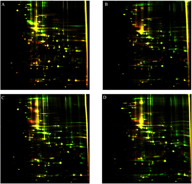
Figure 4. 2D DIGE analysis of various M. gallisepticum isolated from HD3 cells after 7 weeks of infection (Cy3, green) and the laboratory strain M. gallisepticum S6 (Cy5, red).
(A) M. gallisepticum (green). (B) M. gallisepticum initially isolated from HD3 cells (green). (C) M. gallisepticum initially isolated from HeLa cells (green). (D) M. gallisepticum initially isolated from mES cells (green).

