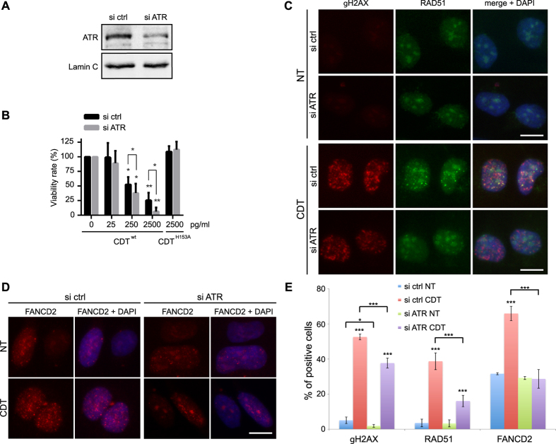
Figure 6. ATR requirement in the signaling and repair of the CDT-mediated DNA damage.
(A) HeLa cells were transfected with scramble (ctrl) or ATR siRNA and soluble cell extracts were prepared to assess the ATR protein level by Western blot analyses. Lamin C is shown as a loading control. Full-length blots are presented in Fig. S8. (B) HeLa cells transfected with scramble (ctrl) or ATR siRNA were exposed for 5 days to CDTwt or CDTH153A and cell viability was analyzed by Crystal violet staining. Results represent the mean ± SD of at least three independent experiments. Statistics were calculated by unpaired Student’s t-test (*P < 0.05; **P < 0.01). (C,D) Representative images of γH2AX and RAD51 (C) or FANCD2 (D) immunostaining in HeLa cells transfected with scramble (ctrl) or ATR siRNA, non-treated (NT) or treated for 24 h with 250 pg/ml of CDTwt. Scale bars = 20 μm. (E) Quantification of HeLa cells transfected with scramble (ctrl) or ATR siRNA positive for γH2AX and RAD51 foci formation from (C) or positive for FANCD2 foci formation from (D). Results represent the mean ± SD of at least three independent experiments. Statistics were calculated by unpaired Student’s t-test (*P < 0.05; ***P < 0.001).

