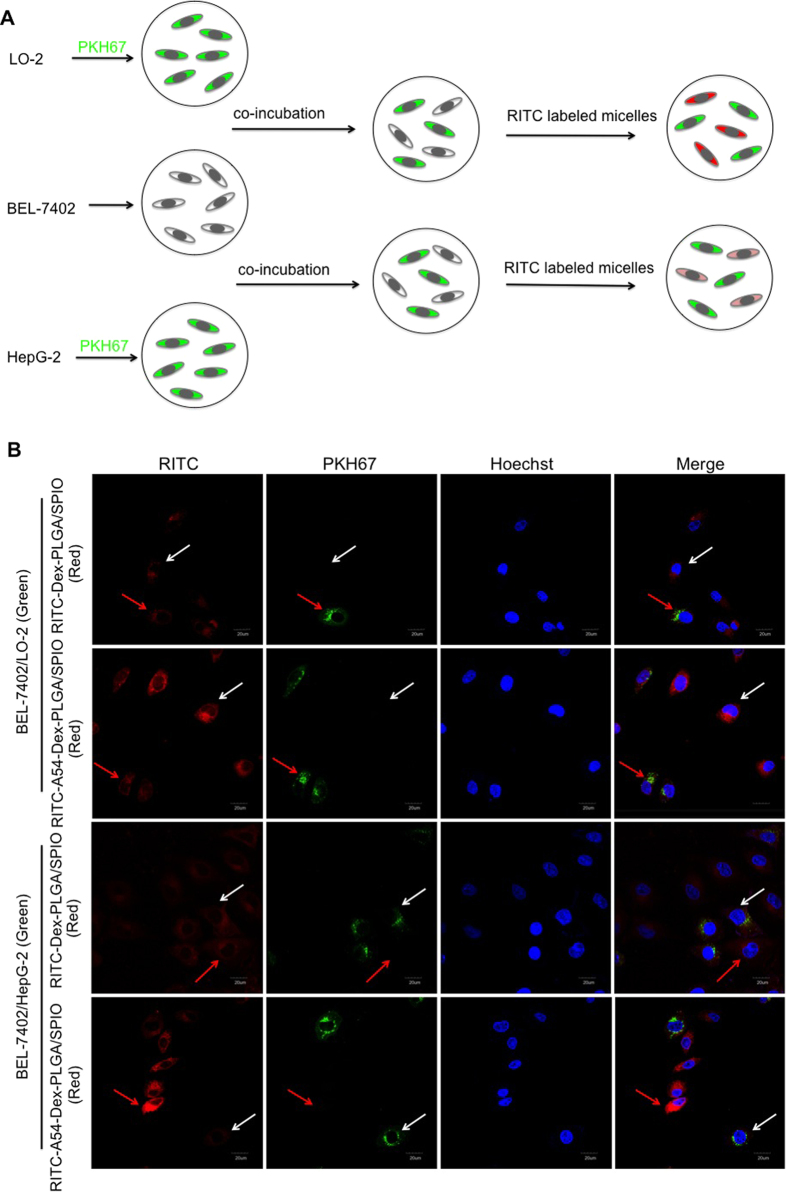
Figure 2.
(A) Schematic diagram of cellular competitive uptake of Dex-PLGA and A54-Dex-PLGA micelles. (B) Confocal microscopy images of RITC labeled micelles for 1 h. HepG2 and LO2 cells (the cytoplasmic membrane labeled with PKH67 fluorescent linker, Green) co-cultured with BEL-7402 cells were incubated with RITC-Dex-PLGA/SPIO and RITC-A54-Dex-PLGA/SPIO micelles (Red). The nucleus were all stained with Hoechst 33342.

