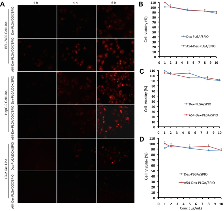
Figure 3.
(A) In vitro cellular uptake of DOX/SPIO-loaded micelles in different cell lines. Fluorescence images of DOX drug after BEL-7402 and HepG2 cells incubated with Dex-PLGA/DOX/SPIO and A54-Dex-PLGA/DOX/SPIO micelles (drug content were 2.5 μg mL−1) for 1, 4, and 6 h, respectively. In vitro cytotoxicity of blank micelles against BEL-7402 (B), HepG2 (C) and LO2 (D) cells. Data represent the mean ± standard deviation (n = 3).

