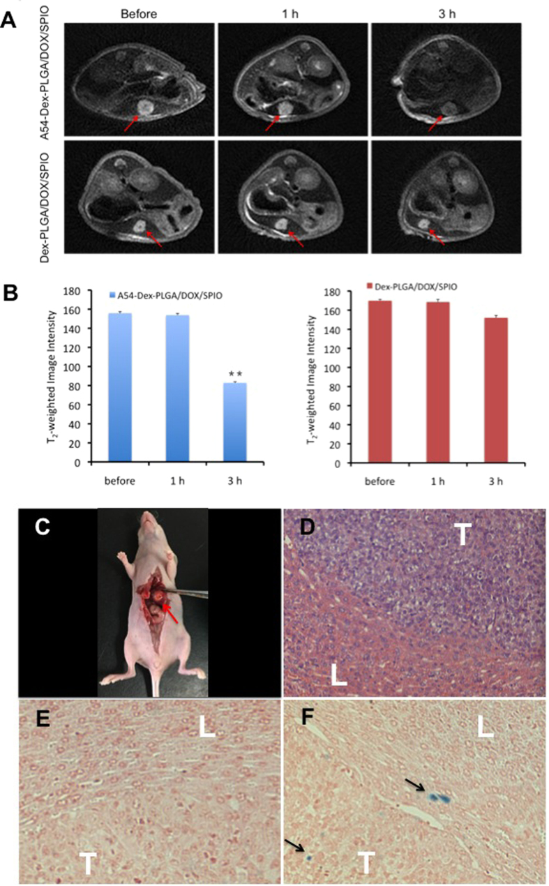
Figure 7.
(A) T2WI images of nude mice bearing BEL-7402 orthotopic implantation tumor before contrast and after injection of Dex-PLGA/DOX/SPIO and A54-Dex-PLGA/DOX/SPIO micelles for 1 h and 3 h, respectively. The red arrow stands for tumors. (B) The T2-weighted image intensity (**p < 0.01). (C) The tumor model made in orthotopic implantation of BEL-7402 tumor. The red arrow stands for the tumor in liver; (D) Microscope image of HE stained live tissue section from nude mice bearing BEL-7402 tumor; Prussian blue stained liver-tumor tissue section of nude mice treated with Dex-PLGA/DOX/SPIO (E) and A54-Dex-PLGA/DOX/SPIO (F), respectively. The black arrow stands for SPIO, “L” stands for the normal liver, “T” stands for the tumor.

