
Figure. Microinfarct and MRI markers of small vessel disease.
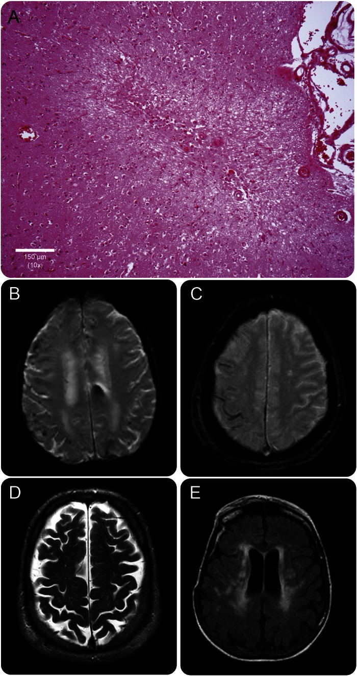
(A) Example of a cerebral microinfarct identified on a cortical section (hematoxylin and eosin stain), (B) lobar microbleeds, and (C) superficial siderosis on T2*-weighted MRI. (D) Dilated centrum semiovale perivascular spaces on T2-weighted MRI. (E) Extensive white matter hyperintensities on fluid-attenuated inversion recovery MRI.