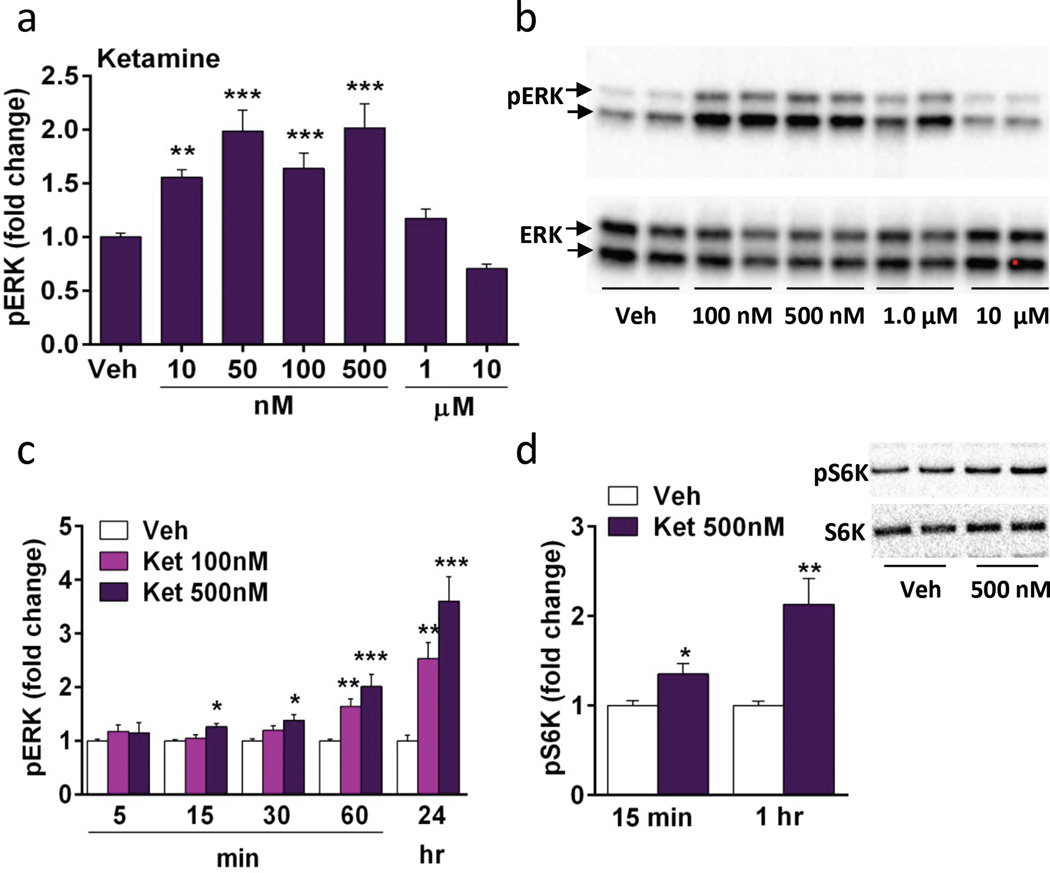
Figure 1. Ketamine incubation increases phospho-ERK and phospho-S6K signaling in a concentration and time dependent manner in vitro.
(a) Following 1 hr of ketamine incubation, there was a concentration dependent increase in phospho-ERK (pERK) with 10–500 nM ketamine. (b) Representative immunoblots for pERK and total ERK. (c) Concentrations of 100 and 500 nM were chosen to test the time course for ketamine stimulation of pERK. (d) Ketamine (500 nM) increases phospho-S6K (pS6K) at 15 and 60 min; representative images of pS6K immunolabeling shown on the right. Levels of total ERK and S6K were determined to control for gel loading and membrane transfer. Results are presented as mean ± SEM. *p < 0.05, **p < 0.005, ***p < 0.0005 compared to vehicle (ANOVA and Fisher’s post-hoc least significant difference test for results in a and c, and Students t test for d).

