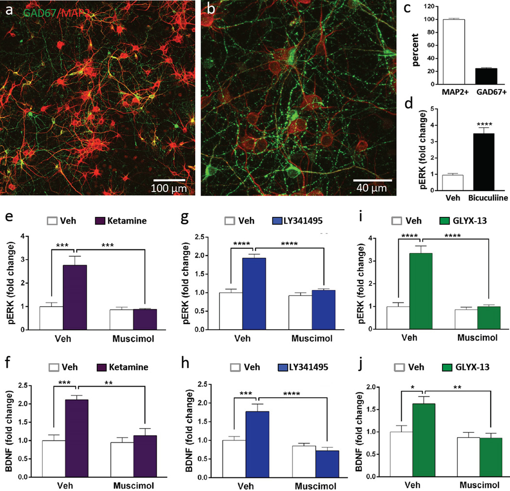
Figure 6. Primary cortical cultures contain GABA interneurons: stimulation of phospho-ERK and BDNF release by rapid-acting antidepressants is blocked by muscimol.
(a, b) Cells plated on coverslips (DIV 10) were fixed and then GAD67 and MAP2 immunolabeling was conducted, demonstrating that a subpopulation of neurons was co-stained for the GABA synthetic marker. Representative low and high power images are shown (scale bars in lower right). (c) The total numbers of GAD67+ cells were determined on 3 separate coverslips and the percentage relative to MAP2+ cells was determined. (d) Incubation with the GABAA receptor antagonist bicuculline (10 µM, 1 hr) significantly increased levels of phospho-ERK (pERK). (e–j) Cultured cells were incubated with the GABAA receptor agonist muscimol (10 µM, 30 min) prior to addition of ketamine, LY341495, or GLYX-13 (1 hr) and levels of pERK were determined by immunoblot analysis (e, g, i), or BDNF release by ELISA (f, h, J). Levels of total ERK and S6K were determined to control for gel loading and membrane transfer. Results are presented as mean ± SEM. *p < 0.05, **p < 0.005, ***p < 0.0005 compared to vehicle or blockade (ANOVA and Fisher’s post-hoc least significant difference test for results).

