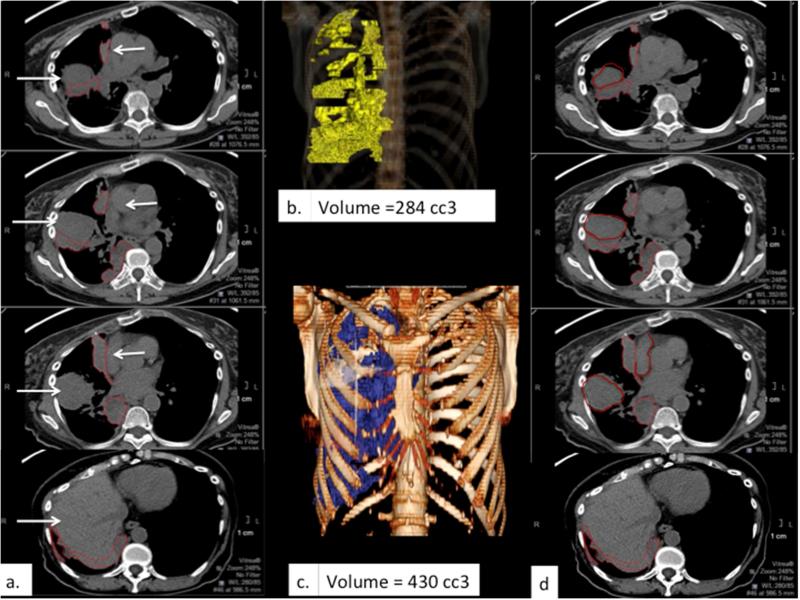
Figure 5.
Example of limited distinction between tumor and adjacent structures. (a) and (d) depict corresponding sets of axial CT images obtained without intravenous contrast in a patient with right-sided MPM, with similar density and therefore poor contrast among tumor, chest wall musculature, liver and the mediastinal vascular structures (white arrows) demonstrating discrepant tumor delineation by the two reference radiologists, respectively. The resulting difference in calculated tumor volume is apparent comparing the respective 3D volume rendered images (b & c).

