Fig. 5.
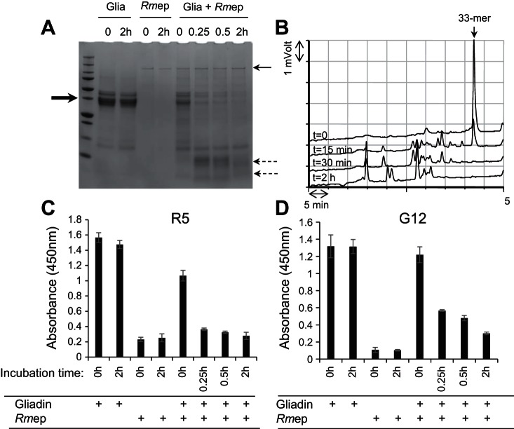
Degradation of mixed gliadins by Rmep and abolishment of immunogenic epitopes. A: gliadins (250 μg/ml) in 50 mM Tris·HCl, pH 8.0, were incubated with Rmep at 57 μg/ml. After time 0 and 15 min, 30 min, and 2 h incubation, 100-μl aliquots were removed, boiled, and analyzed by SDS-PAGE and stained with Coomassie Brilliant Blue. The bold arrow (left) points to the position of the major band in the gliadin preparation, the thin arrow (right) points to the 140-kDa band in the Rmep preparation, and the dashed arrows (right) point to the gliadin degradation fragments. B: reversed-phase (RP)-HPLC of degradation of the immunogenic 33-mer peptide from α-gliadin. Arrow (top) points to the intact 33-mer; C and D: assessment of the survival of epitopes in the gliadin-Rmep degradation mixture determined with the R5 ELISA (C) or G12 ELISA (D).

