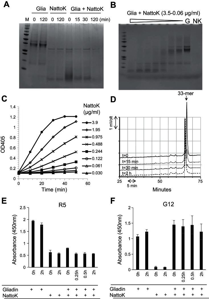
Fig. 7.
Gliadin degradation and epitope abolishment by nattokinase (NattoK) from B. subtilis. A: mixed gliadins (G, 250 μg/ml) were incubated with NattoK (57 μg/ml) for 0, 15, 30, and 120 min. Four lanes on left, controls without enzyme or gliadins, respectively, each at time 0 and 120 min. B: dilution series of NattoK (3.5-0.06 μg/ml) incubated for 30 min with mixed gliadins. Lanes on right, gliadin (G) and NattoK (NK) control. C: dilution series of NattoK incubated with Suc-AAPF-pNA. Hydrolysis was measured at 405 nm. D: RP-HPLC analysis of the gliadin-derived 33-mer (250 μg/ml) incubated for 0, 15, 30, and 120 min with NattoK (57 μg/ml). E and F: epitope abolishment in mixed gliadins (250 μg/ml) incubated for 0, 15, 30, and 120 min with NattoK (57 μg/ml) assessed with the R5 ELISA (E) and G12 ELISA (F).

