Fig. 2.
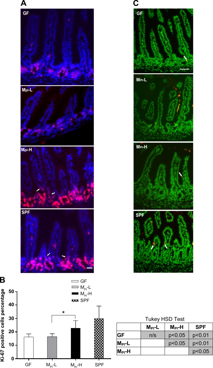
Effect of MPI transfaunation on cell proliferation and apoptosis in ileum. A: immunofluorescence detection of Ki-67 positive cells in ileum of GF, MPI-L, MPI-H, and SPF mice (n = 3–6 mice); arrows refer to the proliferative cells in the lower villus area. B: one-way ANOVA with post hoc Tukey's HSD test was used to compare the groups. *P < 0.05. C: representative TUNEL stained ileum sections in GF, MPI-L, MPI-H, and SPF mice (n = 3–6 mice); arrows refer to the TUNEL positive cells. Bar = 50 μm.

