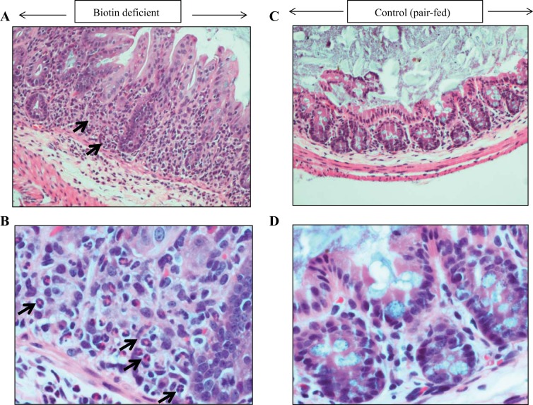
Fig. 6.
Histology of the cecum (A–D) of the biotin-deficient mice and their pair-fed controls. A representative section of cecum of a biotin-deficient mouse (A and B) and its pair-fed control (C and D) is shown with hematoxylin and eosin stain, at ×40 (A and C) and ×200 (B and D). C and D: normal morphology of the pair-fed control cecum. A and B: significant cryptitis and neutrophils within epithelial crypt (arrows).

