Fig. 6.
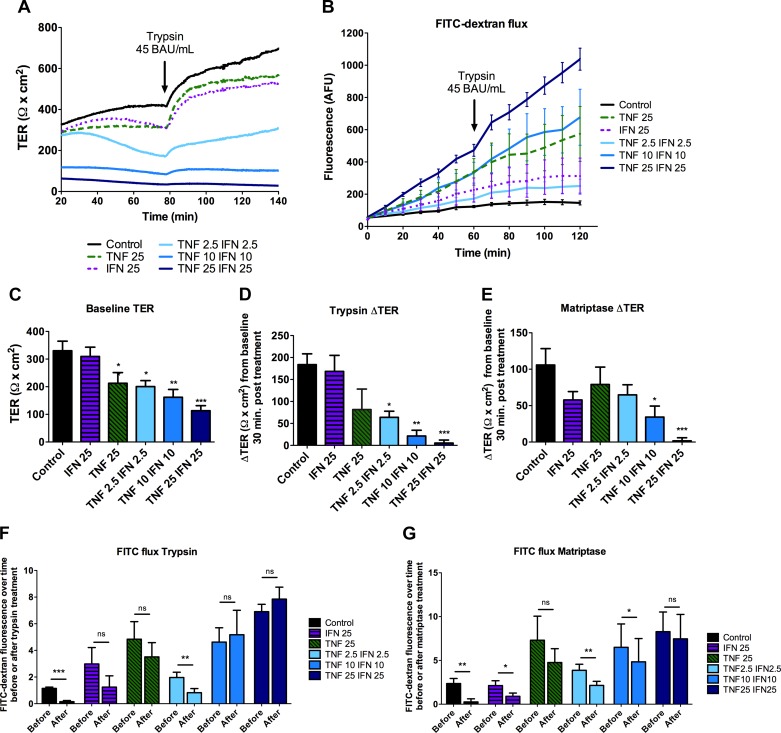
Cytokine-induced barrier disruption of the barrier prevents the serine protease-induced increase in TER. SCBN cells were plated on transwells and grown for 3 days before treating with 2.5, 10, or 25 ng/ml IFNγ and/or TNFα basolaterally every 24 h for 48 h. Cells were mounted in Ussing chambers to assess barrier function. Representative tracing (A) and FITC-dextran flux (B) of cells treated with 45 BAU/ml trypsin (n = 7). Tracings were analyzed for TER before addition of serine proteases (baseline) (n = 13–14) (C), change in TER in response to trypsin 30 min post treatment (n = 5–6) (D), and change in TER in response to 0.5 BAU/ml matriptase (n = 7–8) (E). *P < 0.05, **P < 0.01, ***P < 0.001 compared with untreated control as assessed by ANOVA with Dunnett's post hoc test. To assess change in flux, slopes between 0–60 min and 70–120 min were taken [before or after trypsin (n = 6) (F) or matriptase (n = 4) (G) treatment] and a paired t-test performed within each group. *P < 0.05, **P < 0.01, ***P < 0.001.

