Fig. 6.
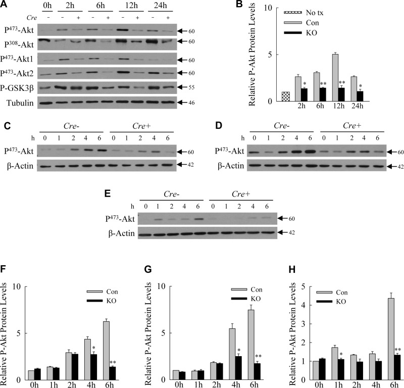
Loss of hepatocyte autophagy is associated with decreased Akt signaling in vivo and in vitro. A: immunoblots of total liver protein from single control (Con) (Cre −) and knockout (KO) (Cre +) mice untreated or treated with LPS for the indicated hours probed for the phosphorylated (P-) forms of Akt and GSK-3β and tubulin. B: relative levels of P473-Akt in Con and KO hepatocytes by densitometric scanning of immunoblots from 3 independent experiments. *P < 0.01, **P < 0.001 compared with Con mice at the same time point; n = 3. C–E: total protein from primary hepatocytes obtained from littermate Con (Cre −) and Atg7Δhep (Cre +) mice and treated in vitro with LPS, TNF, and IL-1β, respectively. Proteins were probed for phosphorylated Akt and β-actin as a loading control. F–H: relative levels of P473-Akt in Con and knockout hepatocytes treated with LPS, TNF, and IL-1β, respectively. *P < 0.01, **P < 0.001 compared with Con mice at the same time point; n = 3. Values are means ± SE.

