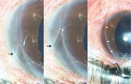
Figure 3. Silicone band leaving an intracorneal groove during removal from the cornea. The free edge of silicone band is marked with black arrows (a,b). Sero-hemorrhagic fluid filling the intrastromal groove after complete removal of silicone band (white arrows) (c).

