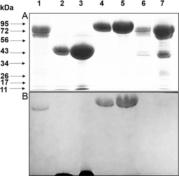
Figure 3.

SDS-PAGE and quinoprotein staining of protein fractions. A. 7.5% gel stained for protein. B. Electroblot which was stained for quinoproteins. A gel identical to that in A was subjected to electrophoretic transfer of the proteins and stained for quinoproteins (see SI for experimental details). Lanes: (1) 20 μg active protein fraction, (2) 20 μg MADH, (3) 80 μg MADH (4) 20 μg LodA, (5) 40 μg LodA (6) 20 μg inactive protein fraction, (7) 100 μg inactive protein fraction.
