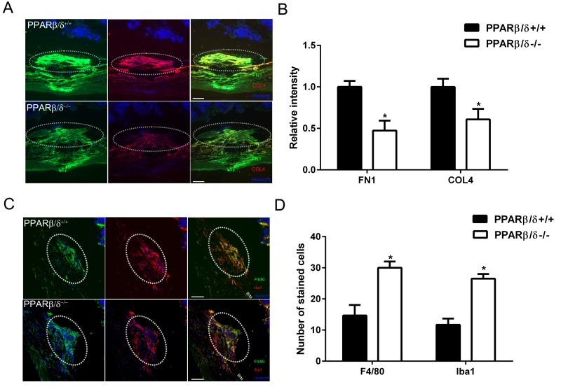
Figure 5. PPARβ/δ regulates extracellular matrix deposition and immune-cell infiltration in CNV lesions.
(A) FN1 (green) and COL4 (red) immunolocalization in CNV lesions of Pparβ/δ+/+ and Pparβ/δ−/− mice (dotted oval demarcates the lesion area; nuclei are stained blue with Hoechst; representative images are shown; scale bar = 50 μm). (B) FN1 and COL4 staining intensity was quantified in the CNV lesions of Pparβ/δ+/+ and Pparβ/δ−/− mice using ImageJ (mean and S.E.M.; n = 3/group; **p < 0.01, two tailed t-test). (C) Laser CNV lesions from Pparβ/δ−/− mice display a higher number of F4/80 (green) and Iba1 (red) immunopositive cells (dotted oval demarcates the lesion area; nuclei are stained blue with Hoechst; representative images are shown; scale bar = 50 μm). (D) The numbers of F4/80+ and Iba1+ cells in the CNV lesions of Pparβ/δ+/+ and Pparβ/δ−/− mice were counted using ImageJ (mean and S.E.M.; n = 3/group; *p < 0.01, two tailed t-test).

